Pos:
Home KnowledgeTechnologyHighly sensitive diamond X-ray detector array for high-temperature applications
X-rays, which have short wavelengths and high energy, have been widely utilized in various fields, such as medical imaging, industrial flaw detection, crystal structure analysis, and security screening2-3. The effectiveness of these applications relies on the availability of high-performance and robust X-ray detectors4. However, the development of detectors that combine high sensitivity, rapid response rates, and stability is a difficult task due to limitations in materials and device detection mechanisms5-6. In this work, a 10×10 X-ray detector array was constructed from single-crystal diamond for the first time. An asymmetric sandwich electrode structure was employed to enhance the detector sensitivity. The diamond X-ray detector array exhibits impressive performance metrics.

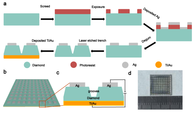
Fig. 1 | Fabrication process of the diamond X-ray detector array. a, Flow chart for the fabrication processes of the diamond detector array. b, Schematic diagram of the diamond detector array. c, Schematic diagram of the diamond detector cells. d, Photograph of the diamond detector array.
The fabrication of the diamond detector array involves several steps, as shown in Fig. 1a. First, 10×10 Ag electrodes are deposited on the front side of the diamond using lithography and magnetron sputtering. Each Ag electrode had a size of 850 μm×850 μm, and the electrode spacing was 120 μm. Next, a Ti/Au common electrode was prepared on the back side of the diamond using magnetron sputtering. To prevent crosstalk between adjacent pixels, trenches are carved between Ag electrodes using laser cutting. A schematic diagram of the diamond detector array is shown in Fig. 1b-c. A photograph of the entire array is shown in Fig. 1d. These trenches effectively suppress the lateral transport of carriers and thus reduce crosstalk between pixels, which is crucial for improving the optoelectronic performance and obtaining clear images of diamond detector arrays.

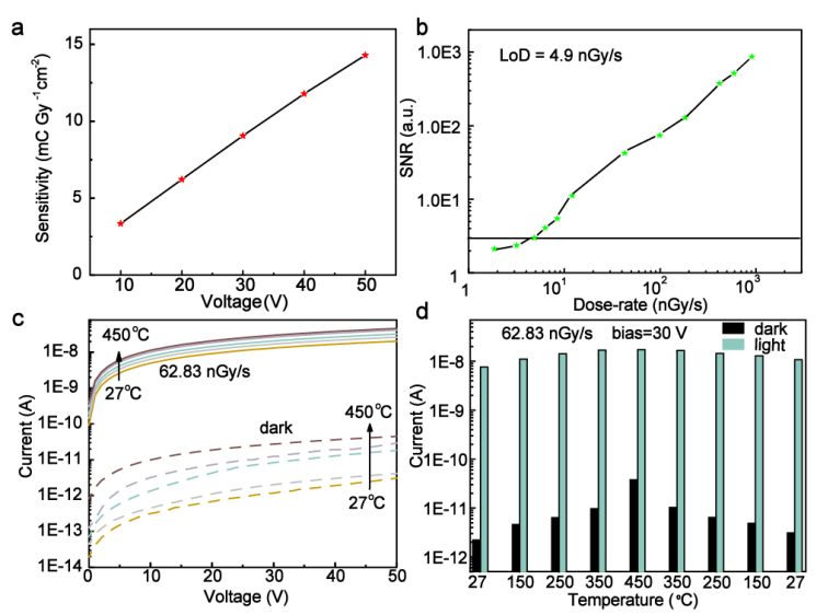
Fig. 2 | Photoelectric properties of the diamond X-ray detectors. a, Sensitivity dependence curves of the detectors at different bias voltages.b, SNR as a function of the dose rate at 40 V bias. c, I–V characteristic curves at different temperatures in the dark and at a dose rate of 32.22 nGy s-1.d, Comparison of light and dark current at different temperatures at a bias of 30 V.
Fig. 2a shows the bias voltage versus response sensitivity curve, showing a sensitivity response of 14.3 mC Gy-1 cm-2 at a bias voltage of 50 V. Fig. 2b shows the signal-to-noise ratio (SNR) with respect to the dose rate, indicating that the detection threshold determined by the intercept of the curve at SNR = 3 is 4.9 nGy s-1. Fig. 2c shows the I-V curves of the detector at temperatures ranging from 27 to 450 °C. It shows that the photocurrent increases continuously with increasing temperature. Fig. 2d compares the photocurrent and dark current with temperature for an x-ray dose rate of 62.83 nGy s-1. Both photocurrent and dark current show good repeatability with increasing and decreasing temperature.

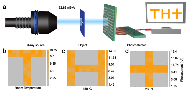
To investigate the imaging capability of the diamond X-ray detector array, an X-ray imaging system is built. The system consists of an X-ray source, a lead-plated masking plate, a diamond photodetector array, and a current collection system, as shown in Fig. 3a. Lead-plated masking plates with hollow patterns of 「T」, 「H」, and 「+」 are placed between the X-ray source and the detector array. The diamond X-ray detector array converts the detected X-rays of different dose rates into electrical signals, and the output currents of the photodetector array are collected using a semiconductor analyzer. The imaging results of the detector array at room temperature, 150 ℃ and 260 ℃ are shown in Fig. 3b-d.
References
1. Dou, W. et al. Highly sensitive diamond X-ray detector array for hightemperature applications.Chip 3, 100106 (2024).
2. Yi, L. et al. X-ray-to-visible light-field detection through pixelated colour conversion. Nature 618, 281-286 (2023).
3. Sakhatskyi, K. et al. Stable perovskite single-crystal X-ray imaging detectors with single-photon sensitivity. Nat. Photonics 17, 510-517 (2023).
4. Chen, J. et al. High-performance X-ray detector based on single-crystal β-Ga2O3:Mg. ACS Appl. Mater. Interfaces 13, 2879-2886 (2021).
5. Bian, Y. et al. Spatially nanoconfined N-type polymer semiconductors for stretchable ultrasensitive X-ray detection. Nat. Commun. 13, 7163 (2022).
6. He, Y. et al. Sensitivity and detection limit of spectroscopic-grade perovskite CsPbBr3 crystal for hard X-ray detection. Adv. Funct. Mater. 32, 24(2022).
https://www.sciencedirect.com/science/article/pii/S2709472324000248
 闽ICP备2021005558号-1
Leave A Message